 History
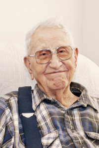
Jim is an 81-year old man who lives with his 80 year old wife, Annie. He has moderate dementia, making him forgetful and unable for example to manage financial matters or help with household chores. He has longstanding urinary incontinence, walks with a zimmer frame and rarely leaves the house.
Jim presented to the Accident and Emergency Unit at his local hospital with a head injury and back pain after a recent fall at home. According to Annie, Jim had become increasingly irritable and confused in the last 36 hours since the fall. She also reported that in the past month, Jim had lost his appetite and had become weaker and more tired.
At the time of examination, Jim was mildly disorientated and confused. His Glasgow coma scale score was 13 and soft tissue swelling and bruising was noted over his forehead, consistent with his fall. Other neurological, cardiovascular and respiratory examinations were normal.
An X-ray of the spine showed a vertebral compression fracture of the thoracic vertebrae at T4 and T5 and, in view of the head injury, Jim was referred for a cranial CT scan. This showed age-related involutional changes and no acute intracranial haemorrhage. However, lucent areas were identified in the skull.
Blood tests showed:
- Haemoglobin level of 78 g/L (range 130 – 170 g/L)
- White cell count 6.2 x 10⁹/L (range 4 – 11 x 109/L)
- Neutrophil count 4.3 x 10⁹/L (range 2.5 – 7.5 x 10⁹/L)
- Platelet count 160 x 109/L (range 150 – 400 x 109/L)
- Calcium 2.95 mmol/L (range 2.2 – 2.6 mmol/L)
- Urea 6.6 mmol/L (range 2.5 – 7.5 mmol/L)
- Creatinine 132mmol/L (range 70 – 150 mmol/L)
- Total protein 83 g/L (60 – 80 g/L)
- Albumin 33 g/L (range 35 – 50 g/L).
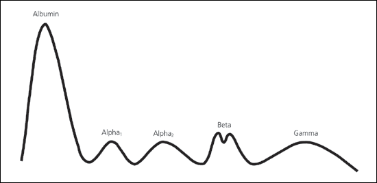
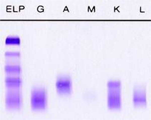
No monoclonal protein was detected in the serum by protein electrophoresis or immunofixation (Figure 1) although there was evidence of immune paresis. Jim’s incontinence made it difficult to collect a urine sample hence a urine protein electrophoresis was not performed.
Jim was managed with intravenous fluids to treat the hypercalcaemia and given a blood transfusion.
Figure 1 – Shows Jim’s SPEP profile (L) and immunofixation (R) – both normal.


